Diagnosis of Urethral Stricture
Diagnosis of Urethral Stricture
It should be done by a urologist to obtain a patient's history and examination.
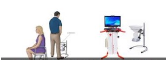
1. Uroflowmetry:
Which is done outpatiently and the patient urinates in the machine that is connected to a computer and processes the information to record the flow rate of the urine, and in the case of the urethral stricture, it will certainly be less than normal.
2. Urthrography:
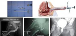
The image of the urethra is done with contrast agent and is one of the most important parts for examining urethral stenosis, which is very important by an experienced technician so that the patient is not harassed and the quality of graphy is acceptable and consists of 2 parts:
- Retrograde urthrography:
A small catheter is placed at the urethra and the contrast agent is injected from it, and the image is taken simultaneously to see the length of the urethral lenght in the graph.
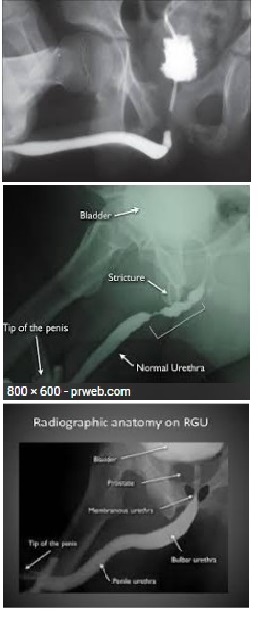
- Voiding cyctourethrography:
The bladder is filled with contrast material and the patient is asked to urinate and the image is taken at the same moment to see the length of the posterior and anterior urethra.
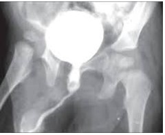
3. Ultrasound of the urethra:
There is no specific preparation for the patient, but it is less likely to be diagnosed.
4. urethroscopy:
That is performed in the operating room by a urologist, and the entire urethra is seen by the phycisian through the bladder area with the endoscopic device.
Some patients need further examination, such as MRI and CT scans, and other tests that the phycisian will consider.

 زبان فارسی